
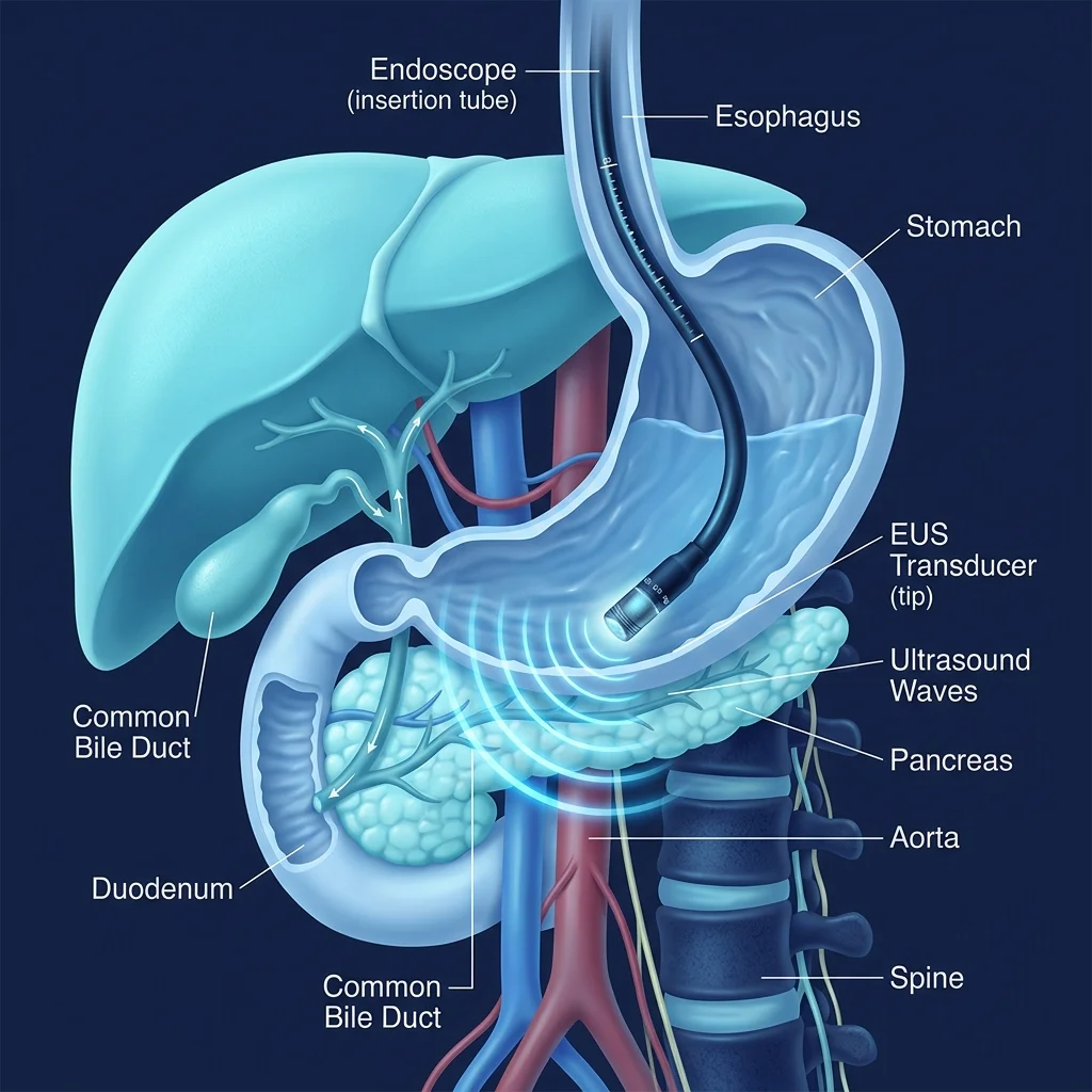
Introduction: When doctors need to examine the stomach, intestine, pancreas, bile ducts, or nearby organs in greater detail, a routine abdominal ultrasound or CT scan may sometimes not provide all the answers.
This is where Endoscopic Ultrasound (EUS) can be extremely useful. EUS is an advanced procedure that combines endoscopy and ultrasound to give doctors a very detailed, close-up picture of the digestive system and the organs lying immediately adjacent to it.
What Is Endoscopic Ultrasound (EUS)?
Endoscopic Ultrasound, commonly called EUS, is a procedure in which a thin, flexible endoscope equipped with a small, high-frequency ultrasound probe at its tip is passed through the mouth into the digestive tract.
The ultrasound probe produces detailed images of organs and tissues that are located just outside the digestive tract wall.
Unlike a conventional ultrasound performed through the skin of the abdomen, the ultrasound probe during EUS is positioned inside the body, very close to the organ being examined. This proximity allows doctors to see fine anatomical structures with exceptional clarity.
What Organs Can Be Examined with EUS?
EUS is particularly valuable when doctors need to investigate deep retroperitoneal structures that can be difficult to evaluate completely with routine abdominal scans. Organs commonly examined include:
Why Might a Doctor Recommend EUS?
Your doctor may recommend EUS when other investigations such as routine ultrasound, CT scan, or MRI/MRCP have not provided a definitive answer. Key clinical indications include:
1. Pancreatic Problems
EUS can provide detailed information about pancreatic cysts, pancreatic masses, chronic pancreatitis, small pancreatic lesions, pancreatic duct abnormalities, and unexplained upper abdominal pain.
2. Detection of Small Bile Duct Stones
Sometimes a patient has severe abdominal pain or abnormal liver blood tests, but standard ultrasound or MRCP does not clearly show a stone. EUS can detect tiny bile duct stones or microscopic sludge (microlithiasis), helping doctors determine whether an interventional ERCP procedure is necessary.
3. Evaluation of Unexplained Jaundice
If a patient develops jaundice (yellowing of eyes and skin) and the root cause remains uncertain, EUS allows detailed examination of the bile ducts, pancreas, and the papilla.
4. Cancer Staging and Evaluation
EUS helps evaluate cancers of the esophagus, stomach, pancreas, and nearby structures. It precisely assesses how deeply a tumor has grown into wall layers and detects whether regional lymph nodes are involved.
5. Tissue Acquisition (Biopsy)
One of the major diagnostic advantages of EUS is enabling the endoscopist to collect tissue samples from deep lesions without open surgery.
How Does EUS Compare to Standard Tests?
Is EUS the same as a normal endoscopy?
No. A routine upper GI endoscopy primarily inspects the surface lining (mucosa) of the esophagus, stomach, and duodenum. EUS combines optical endoscopy with ultrasound, allowing doctors to look beyond the surface lining to examine deeper wall layers and adjacent internal organs.
Is EUS the same as an ultrasound scan?
Not exactly. While both use sound wave technology, a conventional abdominal ultrasound places the probe on external skin. In EUS, the miniature probe is placed directly inside the digestive tract next to the organ, providing far higher resolution image detail.
How Is EUS Performed? Step-by-Step
EUS is performed in an advanced endoscopy suite. The step-by-step process includes:
1. Preparation & Fasting
You will be asked to fast (no food or drink) for 6 to 8 hours before the test to ensure a completely empty stomach.
2. Medical Review
The clinical team reviews your history, blood tests, and current medications (especially blood thinners).
3. Conscious Sedation
Intravenous sedation is administered so that you remain fully relaxed, sleepy, and free of pain during the examination.
4. Scope Insertion
The flexible EUS endoscope is gently passed through the mouth into the esophagus, stomach, and duodenum.
5. Detailed Scanning
The doctor examines the digestive wall and scans surrounding organs like the pancreas, bile ducts, and lymph nodes.
6. Biopsy (If Indicated)
If an abnormality or cyst is found, a very fine needle is guided under real-time ultrasound to sample tissue or fluid.
7. Recovery & Discharge
The scope is removed and you are monitored in the recovery area until sedation wears off (usually within 1-2 hours).
Is EUS Painful, Long, or Risky?
Is EUS painful?
Most patients do not feel pain during EUS because intravenous sedation is given. You may experience a mild sore throat or temporary feeling of bloating afterward, which resolves quickly.
How long does EUS take?
A diagnostic EUS procedure typically takes 30 to 60 minutes. If additional procedures like fine-needle biopsy or therapeutic cyst drainage are performed, it may take longer.
Is EUS safe?
EUS is generally a safe procedure when performed by an experienced endoscopist. Potential risks are low but can include mild bleeding, infection, sedation reaction, tearing (perforation) of the GI tract wall, or temporary pancreatitis (especially after certain pancreatic needle biopsies or interventions).
What Is EUS-Guided Biopsy (FNA / FNB)?
Sometimes imaging scans show an abnormal mass or swollen lymph node, but cannot determine whether it is benign, inflammatory, or malignant. In such situations, tissue sampling is necessary.
During EUS, the specialist passes a specialized microscopic needle through the channel of the endoscope. Under continuous real-time ultrasound monitoring, the needle is directed into the abnormal lesion to safely collect cells or core tissue samples.
- EUS-FNA (Fine Needle Aspiration): Collects fluid or cell clusters for cytological analysis.
- EUS-FNB (Fine Needle Biopsy): Obtains intact tissue cores, enabling detailed histopathological diagnosis and molecular testing.
The collected tissue is sent to the pathology laboratory for accurate diagnosis, helping guide oncology or surgical planning.
Therapeutic EUS: Minimally Invasive Treatments
Yes, EUS is not only diagnostic. With major advancements in interventional endoscopy, EUS is now widely used to perform advanced therapeutic procedures without major open surgery:
1. Drainage of Pancreatic Fluid Collections
Following acute pancreatitis, fluid or necrotic collections (pseudocysts or walled-off pancreatic necrosis / WOPN) can form. EUS allows the specialist to create a safe opening between the fluid pocket and the stomach/intestine under ultrasound guidance, placing lumen-apposing metal stents (LAMS) for internal drainage.
2. EUS-Guided Biliary Drainage
When a bile duct is blocked by cancer or strictures and conventional ERCP fails or is anatomically impossible, EUS-guided biliary drainage provides a safe alternative route to puncture the bile duct from the stomach or intestine and place a drainage stent.
3. EUS-Guided Gallbladder Drainage
In high-risk elderly or critically ill patients with acute cholecystitis who cannot undergo surgical gallbladder removal, EUS can place a stent between the gallbladder and stomach for immediate infection relief.
4. EUS-Guided Pain Relief (Celiac Plexus Block / Neurolysis)
Patients suffering severe, intractable pain from advanced pancreatic cancer or chronic pancreatitis can receive EUS-guided nerve injections (celiac plexus block or neurolysis) to block pain signals and significantly improve quality of life.
5. Tumor-Directed Therapies
Selected specialized centers utilize EUS for radiofrequency ablation (RFA), intratumoral chemotherapy injection, or brachytherapy seed placement directly into targeted lesions.
EUS vs. ERCP: What Is the Difference?
Patients frequently hear about both EUS and ERCP when evaluating bile duct or pancreatic disorders. While complementary, they serve distinct roles:
| Feature | EUS | ERCP |
|---|---|---|
| Primary Role | Detailed diagnostic imaging & tissue sampling | Therapeutic intervention inside duct lumen |
| Modality | High-frequency internal ultrasound scanning | Fluoroscopy (X-ray dye) + side-viewing endoscope |
| Common Uses | Pancreatic masses, staging, small CBD stones, biopsy | Removing CBD stones, widening papilla, stenting |
| Interconnection | EUS often identifies the cause first | ERCP treats ductal blockages subsequently |
Frequently Asked Questions
No. EUS is an advanced investigation and is not required for routine abdominal pain or indigestion. Your doctor recommends EUS when standard blood tests, ultrasound, CT, or MRI scans leave specific diagnostic questions unanswered.
Yes. Because the EUS probe is positioned right next to the pancreas, it is considered one of the most sensitive imaging modalities for detecting small pancreatic lesions and early-stage tumors missed by standard CT scans.
Yes. EUS-guided fine-needle biopsy (FNA/FNB) is performed under continuous real-time ultrasound monitoring, allowing the specialist to avoid blood vessels. The risk of major complications like bleeding or infection is very low (less than 1-2%).
Yes. Most diagnostic EUS and simple needle biopsy procedures are performed on an outpatient basis. After 1 to 2 hours of post-procedure observation, you can return home accompanied by a family member.
Yes. Advanced diagnostic and interventional Endoscopic Ultrasound (EUS) services are available at Siddaganga Hospital, Tumakuru under Dr. Bala Krishna A.
Summary: When to Ask Your Doctor About EUS
Endoscopic ultrasound is a powerful technology that allows doctors to look closely at the digestive tract and surrounding internal organs. It provides vital clarity for pancreatic abnormalities, cysts, small bile duct stones, unexplained jaundice, and suspicious lymph nodes.
If your doctor has advised you to undergo EUS, don't be afraid to ask: "What are we looking for with EUS, and how will the result change my treatment?"
Advanced Interventional Gastroenterology in Tumakuru
Dr. Bala Krishna A. provides state-of-the-art EUS diagnostic and therapeutic interventions at Siddaganga Hospital, Tumakuru.
Dr. Bala Krishna A, MBBS, MD, DM
Consultant Gastroenterologist & Interventional Endoscopist
* This article is intended for general patient education and does not replace medical advice from your treating doctor.